1 东北大学秦皇岛分校控制工程学院,河北 秦皇岛 066004
2 苏州中科行智智能科技有限公司,江苏 苏州 215000
针对合成波长方法动态范围较小的问题,提出一种用于相敏谱域光学相干层析(PSSD-OCT)的大动态范围合成波长相位解包裹方法,解决了传统合成波长方法的相位包裹限制问题,实现了大动态范围、快速及高灵敏度的PSSD-OCT成像。通过选取全长度干涉光谱和位于光谱仪中间的半长度干涉光谱,计算合成相位缺失的周期数;利用全长度干涉光谱及半长度干涉光谱解调结果的分段组合,消除易受噪声影响的相位周期数跳变问题。通过阶差规、硬币及电路板的成像实验,证明该方法可以用于大动态范围(毫米级)的高灵敏度位移解调。
相干光学 相敏谱域光学相干层析 合成波长 相位解包裹 动态范围
Author Affiliations
Abstract
1 School of Control Engineering, Northeastern University at Qinhuangdao, Qinhuangdao 066004, P. R. China
2 Biomedical Engineering Lab, The University of Aizu, Aizu-Wakamatsu, Fukushima 965-8580, Japan
We propose a high-speed all-optic dual-modal system that integrates spectral-domain optical coherence tomography and photoacoustic microscopy (PAM). A 3 × 3 coupler-based interferometer is used to remotely detect the surface vibration caused by photoacoustic (PA) waves. Three outputs of the interferometer are acquired simultaneously with a multi-channel data acquisition card. One channel data with the highest PA signal detection sensitivity is selected for sensitivity compensation. Experiment on the phantom demonstrates that the proposed method can successfully compensate for the loss of intensity caused by sensitivity variation. The imaging speed of the PAM is improved compared to our previous system. The total time to image a sample with 256-256 pixels is -20 s. Using the proposed system, the microvasculature in the mouse auricle is visualized and the blood flow state is accessed.
Photoacoustic microscopy optical coherence tomography angiography dual-modal imaging sensitivity compensation noncontact detection Journal of Innovative Optical Health Sciences
2022, 15(4): 2250023
Author Affiliations
Abstract
1 School of Control Engineering, Northeastern University at Qinhuangdao, Qinhuangdao, Hebei 066004, P. R. China
2 Hebei Key Laboratory of Micro-Nano Precision, Optical Sensing and Measurement Technology, Qinhuangdao, Hebei 066004, P. R. China
3 Department of Ophthalmology, The First Hospital of Qinhuangdao, Qinhuangdao, Hebei 066004, P. R. China
4 Department of Ophthalmology, Qinhuangdao Maternal and Child Health Hospital, Qinhuangdao, Hebei 066004, P. R. China
5 Tangshan Maternal and Children Hospital, Tangshan, Hebei 063000, P. R. China
6 Biomedical Information Engineering Lab, The University of Aizu, Aizu-Wakamatsu, Fukushima 965-8580, Japan
The size and shape of the foveal avascular zone (FAZ) have a strong positive correlation with several vision-threatening retinovascular diseases. The identification, segmentation and analysis of FAZ are of great significance to clinical diagnosis and treatment. We presented an adaptive watershed algorithm to automatically extract FAZ from retinal optical coherence tomography angiography (OCTA) images. For the traditional watershed algorithm, "over-segmentation" is the most common problem. FAZ is often incorrectly divided into multiple regions by redundant "dams". This paper analyzed the relationship between the "dams" length and the maximum inscribed circle radius of FAZ, and proposed an adaptive watershed algorithm to solve the problem of "over-segmentation". Here, 132 healthy retinal images and 50 diabetic retinopathy (DR) images were used to verify the accuracy and stability of the algorithm. Three ophthalmologists were invited to make quantitative and qualitative evaluations on the segmentation results of this algorithm. The quantitative evaluation results show that the correlation coefficients between the automatic and manual segmentation results are 0.945 (in healthy subjects) and 0.927 (in DR patients), respectively. For qualitative evaluation, the percentages of "perfect segmentation" (score of 3) and "good segmentation" (score of 2) are 99.4% (in healthy subjects) and 98.7% (in DR patients), respectively. This work promotes the application of watershed algorithm in FAZ segmentation, making it a useful tool for analyzing and diagnosing eye diseases.
Foveal avascular zone optical coherence tomography angiography watershed algorithm diabetic retinopathy. Journal of Innovative Optical Health Sciences
2022, 15(1): 2242001
Author Affiliations
Abstract
1 School of Computer Science and Engineering Northeastern University Shenyang 110169, P. R. China
2 Shenzhen Academy of Metrology & Quality Inspection Shenzhen 518055, P. R. China
3 School of Control Engineering Northeastern University at Qinhuangdao Qinhuangdao 066004, P. R. China
Segmentation of layers in retinal images obtained by optical coherence tomography (OCT) has become an important clinical tool to diagnose ophthalmic diseases. However, due to the susceptibility to speckle noise and shadow of blood vessels etc., the layer segmentation technology based on a single image still fail to reach a satisfactory level. We propose a combination method of structure interpolation and lateral mean filtering (SI-LMF) to improve the signal-to-noise ratio based on one retinal image. Before performing one-dimensional lateral mean filtering to remove noise, structure interpolation was operated to eliminate thickness fluctuations. Then, we used boundary growth method to identify boundaries. Compared with existing segmentations, the method proposed in this paper requires less data and avoids the influence of microsaccade. The automatic segmentation method was verified on the spectral domain OCT volume images obtained from four normal objects, which successfully identified the boundaries of 10 physiological layers, consistent with the results based on the manual determination.
Optical coherence tomography retinal layers automatic segmentation mean filtering Journal of Innovative Optical Health Sciences
2021, 14(1): 2140011
Author Affiliations
Abstract
1 School of Control Engineering Northeastern University at Qinhuangdao Qinhuangdao 066004, P. R. China
2 School of Computer Science and Engineering Northeastern University Shenyang 110169, P. R. China
Cerebral edema is a severe complication of acute ischemic stroke with high mortality but limited treatment. Although parameters such as brain water content and intracranial pressure may represent the global assessment of edema, optical properties can appear heterogeneously throughout the cerebral tissue relative to the site of injury. In this study, we have monitored the edema formation and progression in both permanent and transient middle cerebral artery occlusion models in rats. Edema was reflected by the decrease of optical attenuation coefficient (OAC) value in OCT system. By utilizing swept-source optical coherence tomography (SS-OCT), we found that in photochemically induced permanent focal stroke model, both the edema size and edema index, steadily developed until the end of monitor (7 h). Comparatively, when transient ischemia was introduced with endothelin-1 (ET-1), the edema was detected as early as 15 min, and began to recover after 30 min until monitor was finished (3 h). Despite the majority of the edema being recovered to some extent, the condition of a small region within the edema kept deteriorating, presumably due to the reperfusion damage which might result in serious clinical outcomes. Our study has compared the edema characteristics from two different acute ischemic stroke situations. This work not only confirms the capability of OCT to temporal and spatial monitor of edema but is also able to locate focal conditions at some areas that might highly determine the prognosis and treatment decisions.
Swept-source optical coherence tomography ischemic stroke cerebral edema optical attenuation coefficient middle cerebral artery occlusion Journal of Innovative Optical Health Sciences
2021, 14(1): 2140006
1 燕山大学电气工程学院, 河北 秦皇岛 066004
2 东北大学秦皇岛分校控制工程学院, 河北 秦皇岛 066004
光学相干层析成像(OCT)技术具有非接触、分辨率高、采集速度快等优势,不仅能够显示样品的三维结构,而且能够检测样品中的运动信息。构建了高分辨率光谱OCT血管成像系统,系统横向分辨率约为6.7 μm,纵向分辨率约为4.7 μm,相机线采集速度140 kHz,能够在2 s内完成三维扫描。在此基础上,通过相邻B扫描图像差分运算提取血流信息,实现血管成像。应用该系统扫描了大鼠大脑皮层的三维血管网络,实验结果表明,系统具有对毛细血管成像的能力。
成像系统 光学相干层析成像 高分辨率 血管成像 在体 激光与光电子学进展
2019, 56(11): 111101
1 东北大学秦皇岛分校实验教育中心, 河北 秦皇岛 066004
2 燕山大学电气工程学院, 河北 秦皇岛 066004
3 东北大学秦皇岛分校控制工程学院, 河北 秦皇岛 066004
提出了一种应用于谱域相位显微成像的相位解包裹方法。利用傅里叶变换及合成波长相位计算方法分别得到具有较小噪声的包裹相位和具有较大噪声的解包裹相位,利用解包裹相位与包裹相位之差计算包裹相位的包裹次数,以此对具有较小噪声的包裹相位进行解包裹。该方法消除了现有方法引入的边界分段错误。建立了一种基于合成波长的谱域相位显微成像系统,使用压电位移台定量验证了该系统可以用于大梯度边界的相位解包裹,并进行了红细胞和倾斜镜面的相位成像。该系统在空气中的位移灵敏度为0.043 nm。
成像系统 谱域相位显微成像 定量相位成像 相位解包裹 合成波长
Author Affiliations
Abstract
1 School of Electrical Engineering, Yanshan University, Qinhuangdao 066004, China
2 School of Control Engineering, Northeastern University at Qinhuangdao, Qinhuangdao 066004, China
3 Experiment Education Center, Northeastern University at Qinhuangdao, Qinhuangdao 066004, China
We demonstrated a method for measurement of central corneal thickness (CCT) with a sub-micrometer sensitivity using a spectral domain optical coherence tomography system without needing a super broad bandwidth light source. By combining the frequency and phase components of Fourier transform, the method is capable of measurement of a large dynamic range with a high sensitivity. Absolute phases are retrieved by comparing the correlations between the detected and simulated interference fringes. The phase unwrapping ability of the present method was quantitatively tested by measuring the displacement of a piezo linear stage. The human CCTs of six volunteers were measured to verify its clinical application. It provides a potential tool for clinical diagnosis and research applications in ophthalmology.
170.4500 Optical coherence tomography 170.4460 Ophthalmic optics and devices Chinese Optics Letters
2019, 17(4): 041701

Author Affiliations
Abstract
1 School of Control Engineering, Northeastern University at Qinhuangdao, Qinhuangdao 066004, China
2 School of Electrical Engineering, Yanshan University, Qinhuangdao 066004, China
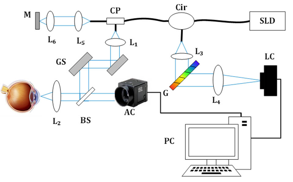
We demonstrate a system for measuring the ocular axial length (AL) with high sensitivity and high speed using spectral-domain low-coherence interferometry (SD-LCI). To address the limit in measuring such a large range by using SD-LCI, we propose a full-range method to recognize the positive and negative depths. The reference arm length is changed synchronously with the shift of the focal point of the probing beam. The system provides a composite depth range that is sufficient to cover the whole eye. We demonstrate the performance of the presented system by measuring the ALs of five volunteers. This system can provide the A-scan ocular biometric assessment of the corneal thickness and AL in 0.1 s.
170.4500 Optical coherence tomography 170.4460 Ophthalmic optics and devices Chinese Optics Letters
2018, 16(3): 031701
1 东北大学秦皇岛分校控制工程学院, 河北 秦皇岛 066004
2 东北大学秦皇岛分校实验教育中心, 河北 秦皇岛 066004
采用光学相干层析研究了粒子流背向散射光强的涨落特性,将粒子对背向散射光的影响分为相位调制和振幅调制两部分,建立了粒子流流速全部分量测量方法,由背向散射光强信号分别得到粒子流多普勒频移和渡越时间,进而计算出流速的纵向分量和横向分量,用聚苯乙烯粒子悬浮液对这种流速测量方法进行了实验验证。
医用光学 光学相干层析 流速 多普勒频移 自相关 光学学报
2014, 34(11): 1117002


